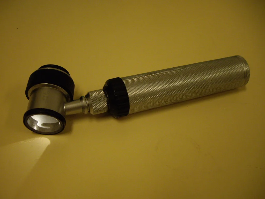

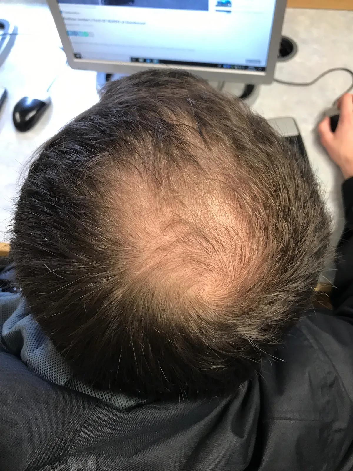
Trichoscopy, the use of a handheld dermatoscope to examine the scalp and hair shafts at magnification of typically 20–70×, has become the standard diagnostic tool in dermatology hair clinics over the past fifteen years. It enables visualisation of follicular openings, perifollicular structures, hair shaft characteristics, and scalp surface features that are invisible to the naked eye. For a tool that costs a few hundred pounds and adds minutes to a consultation, the diagnostic uplift is enormous.
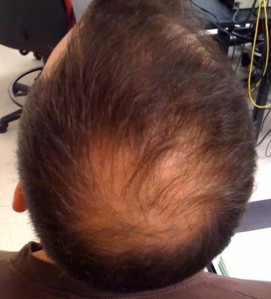
Specific trichoscopic features differentiate hair loss types. Androgenetic alopecia shows hair shaft diameter variability (different thicknesses in different follicles), peripilar brown halos around miniaturised follicles, and an increased number of vellus hairs. Alopecia areata shows characteristic 'exclamation mark' hairs, yellow dots representing empty follicles, and black dots from fractured hairs. Scarring alopecias show loss of follicular openings, the most important single finding because it indicates permanent destruction.
Despite the diagnostic value, many primary care consultations and even some dermatology consultations still rely on visual inspection alone. Patients are reasonable to request trichoscopy if they're paying for a hair loss consultation, the absence of it suggests the practitioner is missing the standard diagnostic tool of contemporary hair medicine. Trichoscopy also enables objective monitoring of treatment response, with serial measurements of vellus-to-terminal hair ratios providing quantitative tracking that visual inspection cannot deliver.
Discussion (3)
DrewFromAustin
4 months ago
Anyone tried this in combination with low-dose oral minoxidil? Wondering if mechanisms stack.
Priya S.
4 months ago
The references section is what makes this site worth reading, actual PubMed links, not affiliate-stuffed nonsense.
James_NW3
4 months ago
Reasonable take. I'd still want to see longer follow-up before drawing strong conclusions.
Join the discussion
Free account. Read, like, save, and comment on every article.