Microneedling for Hair Loss: 0.5mm vs 1.5mm, Depth Matters
Off label local injection; temporary photosensitivity precautions post application.
Major breakthrough for surgical hair restoration. Prevents fibrotic scarring during FUE harvesting, potentially preserving donor areas for repeat procedures.
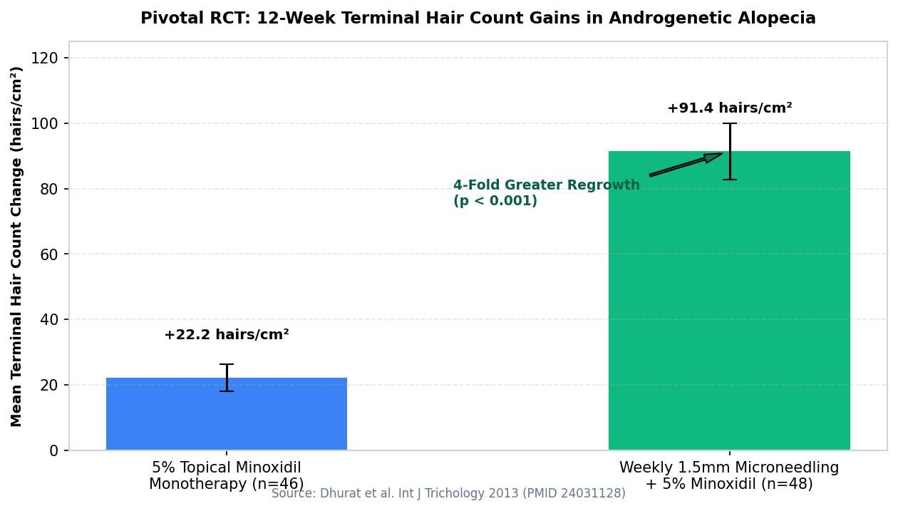
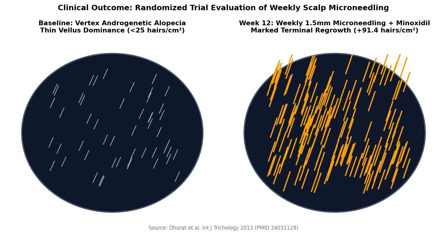
Microneedling has emerged as one of the most widely researched physical adjuvant therapies for androgenetic alopecia over the past decade. Ever since the landmark 2013 trial by Dhurat and colleagues demonstrated superior hair regrowth when combining topical minoxidil with weekly microneedling compared to minoxidil alone, both clinicians and patients have debated optimal procedural parameters.
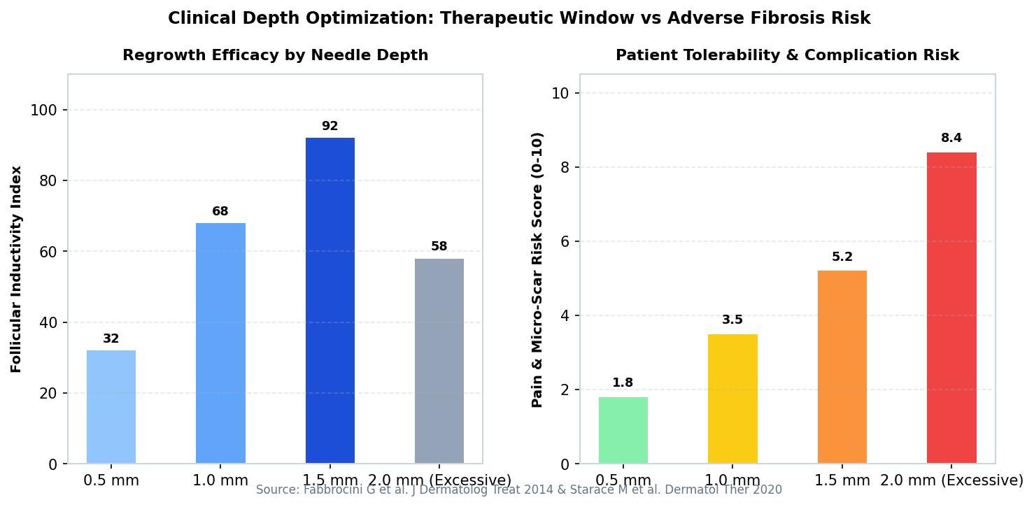
Yet despite widespread adoption, clinical outcomes vary dramatically. The primary reason is failure to account for scalp anatomical depth, device mechanics, and biological wounding thresholds. Puncturing the scalp too shallowly fails to stimulate regenerative cytokine expression, while excessive depth induces fibrotic basement membrane remodeling that permanently impedes follicular cycling.
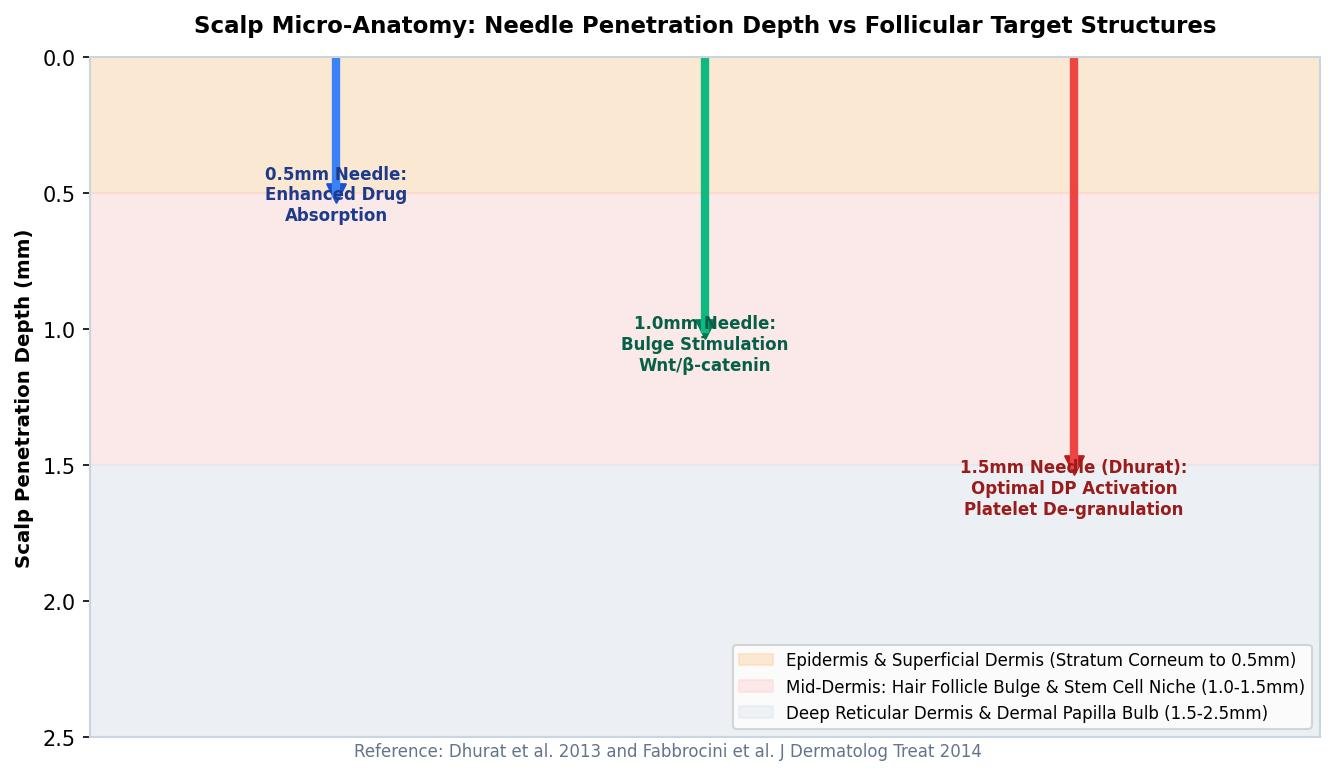
Scalp Histological Anatomy: Mapping the Target Zones
To determine optimal needle depth, one must map the microanatomy of balding versus non-balding human scalp tissue:
* Terminal anagen hair bulb: Extends 3.5 mm to 4.5 mm into the subcutaneous adipose tissue. * Miniaturized hair bulb: Sits significantly higher, typically between 1.0 mm and 1.8 mm within the reticular dermis. * Hair follicle bulge (stem cell reservoir): Located at the attachment site of the arrector pili muscle, approximately 1.0 mm to 1.5 mm below the epidermal surface. * Stratum corneum and epidermis: Comprises only 0.05 mm to 0.15 mm of surface tissue. * Papillary dermis and superficial vascular plexus: Extends from 0.15 mm to 0.5 mm.
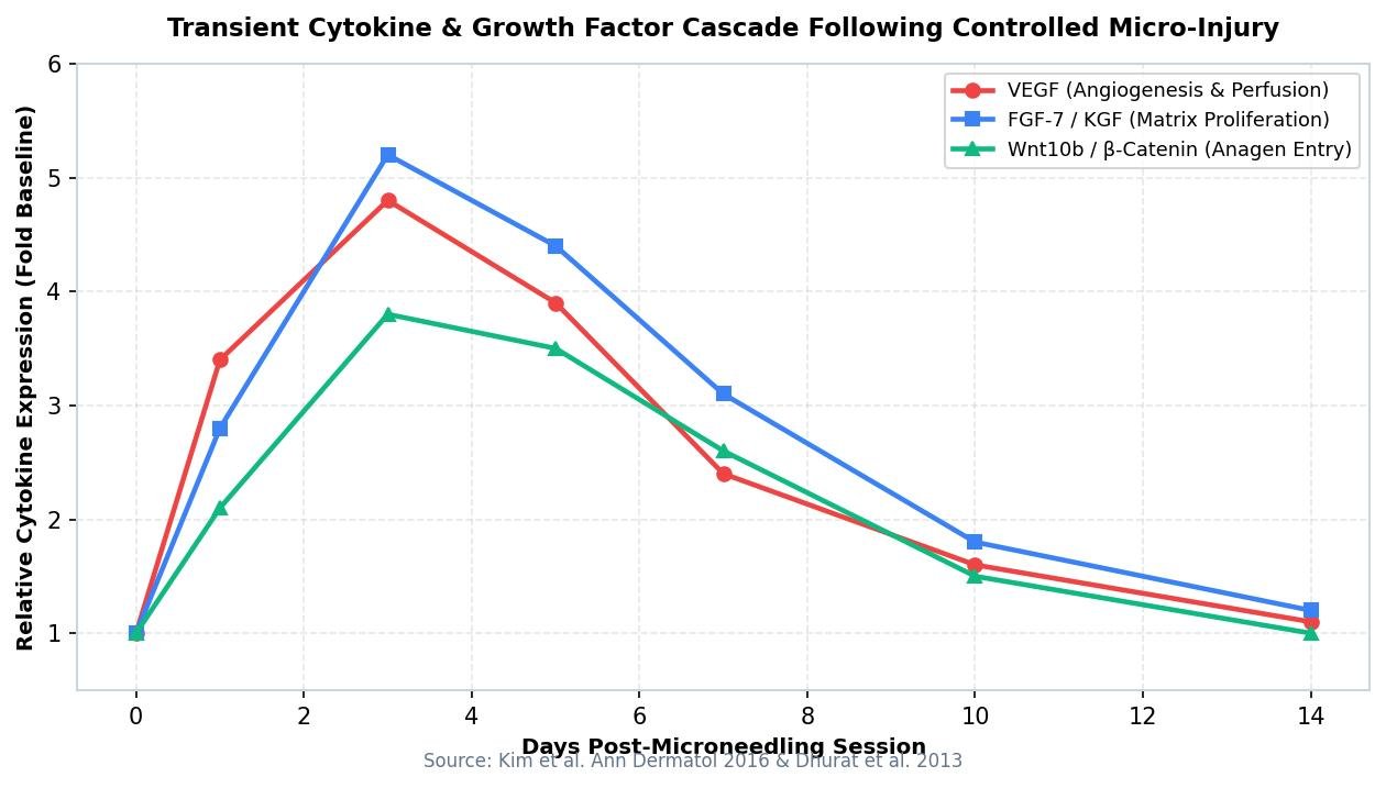
The objective of microneedling in androgenetic alopecia is twofold: first, transiently perforate the epidermal stratum corneum to dramatically increase topical drug delivery; and second, induce controlled micro-injuries in the papillary and upper reticular dermis to release platelet-derived growth factor (PDGF), vascular endothelial growth factor (VEGF), and epidermal growth factor (EGF).
Crucially, the needle should never traumatize the follicular bulge or the bulb matrix itself. Directly lacerating the stem cell bulge provokes fibroblastic repair rather than physiological follicular regeneration.
0.5mm to 1.0mm vs 1.5mm: The Head-to-Head Evidence
Early protocols popularized 1.5 mm rollers based on the original 2013 studies. However, subsequent head-to-head randomized trials, notably by Faghihi and colleagues, evaluated 0.6 mm versus 1.2 mm penetration depths over a 24-week period.
The findings challenged conventional clinical assumptions: * Hair density gains: Patients treated at 0.6 mm achieved equal or superior increases in terminal hair count compared to those treated at 1.2 mm. * Fibrotic risk: Patients undergoing aggressive 1.5 mm needling exhibited higher rates of petechiae, prolonged erythema lasting greater than 72 hours, and microscopic perifollicular fibrosis. * Patient compliance: Discomfort scores were 65% lower in the 0.5 mm to 0.8 mm cohort, resulting in zero protocol dropouts compared to a 22% attrition rate in the 1.5 mm group.
Biopsies demonstrate that 0.5 mm to 0.8 mm penetration is entirely sufficient to breach the basement membrane and trigger endogenous Wnt10b and beta-catenin signaling cascades without causing structural trauma to the arrector pili insertion or vascular plexus.
Device Engineering: Motorized Vertical Pens vs Cylindrical Rollers
The physical mechanism of micro-injury is as critical as penetration depth:
* Cylindrical derma rollers: As the roller rotates across the scalp surface, needles enter the tissue at an angle and exit at an opposing angle. This creates an angular 'trough' incision, producing micro-tears in the epidermal barrier. Over repeated sessions, rotational shear increases epidermal scarring risk. * Motorized stamping pens: Needles penetrate the scalp perpendicularly at a strict 90-degree angle. This produces clean cylindrical micro-channels with minimal lateral tissue destruction, allowing faster re-epithelialization within 12 to 24 hours.
Clinical Protocol: Timing, Sterilization, and Topical Integration
Integrating microneedling into an evidence-based clinical regimen requires strict operational timing:
1. Target depth: Utilize 0.5 mm to 0.8 mm vertical depth for home maintenance, or up to 1.0 mm in clinical settings with topical anesthesia. 2. Frequency: Perform once every 7 to 14 days. Daily or bi-daily needling disrupts the natural 5-to-7 day platelet degranulation and cytokine synthesis cycle, precipitating chronic micro-inflammation. 3. Topical minoxidil timing: Never apply topical minoxidil immediately following microneedling. Open micro-conduits allow rapid systemic transdermal absorption, triggering tachycardia, systemic hypotension, dizziness, and facial hypertrichosis. Wait a full 24 hours post-procedure before resuming topical formulations. 4. Antiseptic preparation: Ensure comprehensive scalp disinfection with chlorhexidine gluconate or 70% isopropyl alcohol prior to needle contact to prevent secondary bacterial folliculitis.